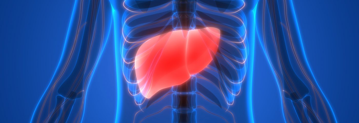
Diaphragmatic endometriosis is rare but may lead to a rupture of the diaphragm, the muscle essential for breathing, without any or very subtle symptoms, a case report and literature review details.
The report, “Non-traumatic diaphragmatic rupture with liver herniation due to endometriosis: A rare evolution of the disease requiring multidisciplinary management,“ describes the case of a women whose diaphragm tore due to the growth of endometrial lesions, causing part of her liver to move upward into the chest (liver herniation). It was published in the Journal of Gynecology Obstetrics and Human Reproduction.
Diaphragmatic endometriosis usually involves the right and tendon part of the diaphragm, and doctors should be aware that symptoms of a rupture are subtle — normally intermittent pain in the right thorax or catamenial pneumothorax, which is a collapse of the lungs due to air leakage into the space surrounding the lungs. A magnetic resonance imaging (MRI) scan is the exam of choice to confirm this complication, which requires surgery for treatment.
Thoracic endometriosis is a rare condition in which endometrial tissue — tissue from the inner lining of the uterus (called the endometrium) — becomes displaced and attaches to areas in or around the lungs.
The 35-year-old woman in this case had a form of thoracic endometriosis that affected the diaphragm, the muscle at the bottom of the chest essential for breathing.
She had a medical history of stage 4 pelvic endometriosis (diagnosed in 2011) and was referred in 2017 to the Caen University Hospital in France, complaining of right shoulder pain that worsened with menstruation and was associated with chronic pelvic pain.
A computed tomography (CT) scan confirmed she had a diaphragm rupture and a liver hernia — a portion of her liver had moved upward through the diaphragm opening into the space that surrounds the lungs (pleural cavity). Moreover, there were patches of tissue on the hernia’s edges, later confirmed by MRI to be endometrial tissue.
The MRI also revealed other endometriotic lesions: an ovarian cyst, bleeding into one of the fallopian tubes, and nodules in the uterus.
To treat her, doctors opted for a muscle-sparing mini-thoracotomy, a surgery in which a cut is made between the ribs to reach the lungs or thorax. During the procedure, the displaced portion of the liver was put back into the abdominal cavity, endometriotic lesions were removed, and the diaphragm opening was closed. Due to the large defect, which was worsened by the resection of lesions, an absorbable mesh also was placed to strengthen the diaphragm.
The patient recovered well, and a biopsy of the removed lesions was consistent with endometrial tissue. Six months after surgery, her chest X-ray looked normal, and she did not have any more shoulder pain.
Hormonal therapy with a contraceptive pill was prescribed as she refused to take GnRh agonists, worried about possible side effects.
In addition to this case, the researchers found 12 other reports in the literature of diaphragmatic rupture caused by endometriosis. They all share many similarities, including subtle symptoms, which happen in 30% of the cases. Most commonly, these are pain in the right thorax or recurrent cathamenial pneumothorax.
“Symptoms are rather subtle and do not foreshadow the importance of diaphragmatic defect,” the researchers said.
These signs require imaging exams, according to the researchers, who said that “MRI is the key examination to diagnose rupture.”
The cause of this type endometriosis is not yet known. Pelvic endometriosis may spread into the diaphragm over the course of several years, either due to menstrual reflux or passage through spaces between the colon and the abdominal wall.
Diaphragm ruptures associated with endometriosis have characteristic marks: they are typically right-sided, central in position, and located in the tendon part of the muscle, according to the researchers.
In contrast, spontaneous rupture of the diaphragm not linked with endometriosis, due to fragility or malformation, can be left-sided and happens mostly at the diaphragm margins.
“A multidisciplinary approach is necessary to choose the best therapeutic option,” the researchers emphasized.
In case of rupture, a surgery is always necessary, if possible through a laparoscopy (keyhole surgery), which is less invasive than the thoracotomy performed in this study.
However, a thoracotomy “might be useful in cases of complex adherences between the diaphragm or lung and herniated organs,” the researchers added.

