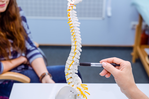
Damage to the nerve fibers that link the spinal cord to the pelvic region, called the sacral nerve, may help explain chronic pain associated with endometriosis, a study has found.
The findings were reported in the study, “The Role of MRI-DTI in Predicting Pain Related to Endometriosis: a Preliminary Study,” which appeared in The Journal of Minimally Invasive Gynecology.
Chronic pain is a symptom shared by many women who have endometriosis. Painful periods, painful sexual intercourse, and chronic pelvic pain are the most common pain symptoms these women experience. In rarer cases, painful urination or defecation also can occur.
Although seen as a common symptom of endometriosis, the cause of this pain is not completely understood. Several factors have been reported to be associated with endometriosis-associated pain, including inflammation, hypersensitivity, oxidative stress, and anatomic distortion, among others.
The underlying process of endometriosis comprises a complex network of cellular signals and tissue differentiation. Studies have suggested that pain symptoms could be due to compression or infiltration of nerve fibers by adhesive endometrium lesions. Also, signals promoting the growth of nerve cells close to the lesions may contribute to the development of pain.
In a previous study, a team of researchers at “Sapienza” University of Rome showed that women with endometriosis and chronic pain presented significant changes in the structure of sacral nerve roots compared to healthy controls.
To better understand the association between sacral nerve root damage, pain symptoms, and endometriosis surgical findings in women, the team in the most recent study evaluated sacral nerve damage in 76 women suspected of having endometriosis.
The nerve analysis used combined magnetic resonance imaging (MRI) and diffusion tensor imaging (DTI), which allowed researchers to virtually recreate the three-dimensional microstructures of nerve fibers.
The sacral nerve MRI-DTI analysis revealed that sacral roots were found to be normal in 17 patients, while 44 women showed alterations in the architecture of the nerve roots. These changed nerve fibers were found to have irregular and disorganized structures, with a loss of simple, unidirectional course.
After surgical evaluation and treatment, the researchers found ovarian endometriomas in 82.1% of patients and deep lesions in 57.1%.
Analysis of all the information revealed that changes in the sacral nerve structure detected by MRI-DTI were significantly associated with the severity of menstrual pain and the duration of chronic pelvic pain. Additionally, they found that women who had deep lesions had 23.32 times higher risk of having sacral nerve root damage. Patients who had tubo-ovarian and cul-de-sac adhesions had 8.1 times higher risk.
Overall, these findings suggest that damage to the structure of the sacral nerve could be related to deep lesions and the development of endometriosis, and could in part explain pain symptoms in these patients.
The study also shows that use of MRI-DTI not only can improve the understanding of endometriosis-associated pain, but it may also help identify patients that could benefit from targeted and more efficient pain therapies.
“We hope that DTI may in the future become useful in the planning of a more personalized therapeutic approach by selecting those patients who may possibly benefit from different treatments,” the researchers said.

