In a new study entitled “A Tissue Specific Magnetic Resonance Contrast Agent, Gd-AMH, for Diagnosis of Stromal Endometriosis Lesions: A Phase I Study,” researchers evaluated the expression of anti-mullerian hormone (AMH) as a potential cellular marker of endometriosis lesions in vivo. The study was published in the Journal of Cellular Physiology.
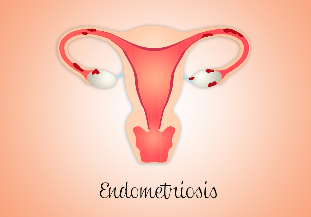
Endometriosis, a benign disease characterized by the presence of endometrial tissue outside the uterus, is one of the most common gynecological disorders affecting 10% of women of reproductive age and increasing to 50% in women with fertility issues. Endometriosis diagnosis is performed by surgical procedures — laporoscopy or laparotomy — both requiring abdominal incision to gain access into female pelvic organs, and it requires the visual confirmation of endometriosis lesions. Frequently, however, endometriosis lesions have very reduced sizes rendering its precise location impossible to pinpoint with standard methods.
In this new study, authors evaluated the expression of anti-mullerian hormone (AMH) in adult human and endometriosis tissues, whose presence in endometrium and endometriosis lesions was suggested in previous studies, and determined whether it could be used as a marker for endometriosis lesions in vivo. To this end, the team analyzed first the expression of AMH in a wide collection of adult human tissues, and found AMH is ubiquitously expressed throughout human organs and tissues. Particularly high expression of AMH levels were detected in the female genital system, notably in endometriosis lesions, as previously shown. Using an antibody against AMH, the team showed that indeed they could detect small endometriosis lesions (5 mm in diameter) by magnetic resonance in an in vivo (mouse) model.
Thus, authors note that their results suggest the potential of AMH as an efficient marker of AMH can be used as an effective cellular target to specific location in vivo, even in small foci of endometriosis. Additionally, AMH could be used to detect the existence of residual lesions after surgical removal. Notably, the team observed no toxicity or behavioral changes in mice treated with anti-AMH labeled with gadiolinium (Gd). While additional studies are still required to further evaluate and establish the exact protocol for Gd-AMH in magnetic resonance studies, this first study established the potential of AMF as a diagnosing strategy for in vivo endometriosis and the extent of endometriosis lesions.

