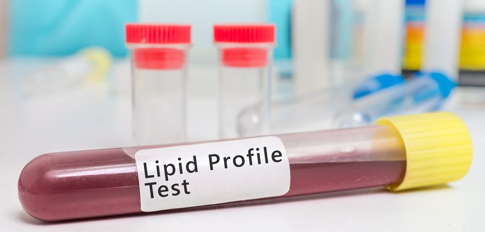
Pennsylvania State University researchers have revealed that lipid metabolism, a parameter easily measured with a blood test, is different in mice models of endometriosis. The finding may ultimately lead to a new diagnosing tool for endometriosis patients.
The study,”Metabolomics revelas altered lipid metabolism in a mouse model of endometriosis,” was published in the Journal of Proteome Research.
Currently, laparoscopic surgery is the standard method used to diagnose endometriosis disease. The invasive procedure requires inserting a tube equipped with a tiny video camera into the peritoneal cavity so that physicians can see the involved organs.
It is believed that environmental conditions of the peritoneal cavity, including chronic inflammation, help endometrial cells proliferate. Now, the recent studies shows that the disease may be associated with altered lipid signaling.
The research team led by Mainak Dutta and Andrew D. Patterson, induced endometriosis in mice and then analyzed them under a mass spectrometry-based lipidomics approach in order to seek alterations in the mice serum lipid profiles.
Dysregulated lipids were found in the animals; triglycerides (TGs) and phosphatidylethanolamines (PEs) were decreased, whereas sphingomyelins (SMs) and phosphatidylcholines (PCs) were upregulated in mice with endometriosis. However, TGs were also decreased in a mouse model of peritoneal inflammation, suggesting that TG may be associated with inflammation but not endometriosis.
The also found that PC and PE ratio in the serum of the mice with endometriosis was altered, suggesting that a specific lipid pathway was activated most likely as a consequence of the abnormal estrogen metabolism associated with the disease.
This study not only provides new insight on the etiology of endometriosis but it may also help develop new ways of diagnosing patients by measuring serum lipid levels, particularly the PC and PE ratio.
The study authors concluded: “Though the expression of a large number of lipids was altered in endometriosis mice, a number of them appear to be due to the peritoneal inflammation associated with the disease. The results of PC and PE expression seem promising and can thus be further validated in clinical samples for its possible use as a marker for endometriosis.”

